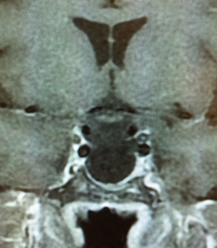
From Lewis Blevins, MD – Empty sella syndrome as a misnomer for the sella is not empty. It is usually filled with spinal fluid. Furthermore, a true syndrome does not exist. An empty sella is a radiological finding. Sometimes, this is of clinical importance and at other times it is not.
The sella turcica is the saddle-shaped hollowing, or fossa, in the sphenoid bone that houses the pituitary gland. The roof of the sella is covered by a reflection of the dura mater known as the diaphragma sellae. There is usually a small opening in the diaphragma sellae to permit the pituitary stalk and associated portal blood vessels to pass from the hypothalamus down into the pituitary fossa.
An empty sella may be primary or secondary in nature. Primary implies that one was born with the likelihood of developing this abnormality. In these patients, the small opening in the diaphragma sellae is larger than usual because the dural reflection did not develop properly. This allows the pressure of the spinal fluid to cause herniation of the arachnoid membranes through the hole into the sella. As a result, the pituitary gland is flattened in the floor of the sella and the pituitary stalk goes straight to the glandular tissue. This occurrence produces what is known as the “sword sign” because the pituitary stalk looks like a sword hanging downwards from the hypothalamus. A secondary empty sella is due to some underlying disease process that causes destruction of the pituitary gland. In many cases, the sella is larger than normal whereas it is of normal size in those with primary empty sella. The common causes include Sheehan syndrome and the end-stage of lymphocytic hypophysitis as well as necrosis of the pituitary tumor or surgery or some other pituitary disease process. For example, I have seen patients to look acromegalic but have an enlarged empty sella. The hypothesis is that they once had a large pituitary tumor that degenerated and was ultimately resorbed. Intrasellar arachnoid cysts can often be mistaken for an empty sella as can large Rathke’s cleft cysts. In most of these cases, there is no sword sign and the pituitary stalk is deviated to one side or the other. Of course, these patients with cystic lesions may require surgical intervention were as surgery is not performed in those patients with a traditional empty sella. Lastly, an empty sella can be associated with pseudotumor cerebri.
The most important determinations when faced with empty sella are: is this disorder primary or secondary in nature; if secondary in nature is therapy for an underlying disorder required; regardless of cause, as the pituitary functioning normally; if there is pituitary dysfunction what hormone replacement is required.
Patients who have a primary empty sella usually have normal pituitary function. However, some, in my experience about 25%, have isolated growth hormone deficiency. Some have mild hyperprolactinemia. Some of these patients have a coexisting microadenoma of the pituitary gland that produces prolactin. A rare patient will also have mild central hypothyroidism or irregular menses. DI is rare in primary empty sells and suggests an underlying disease process rather than a developmental abnormality. Patients with a secondary empty sella may have one or more pituitary deficits with more than half having growth hormone deficiency. Hormone replacement is straightforward according to principles and practices with all of the caveats employed in patients who have hypopituitarism. Rarely, the optic pathways will sink down into the empty sella causing visual deficits and these patient’s require surgery to perform a chiasmapexy where a lump of fat is put into the sella to hold up the visual pathways and restore vision to normal.
© 2014, Pituitary World News. All rights reserved.
