From J D Faccinetti Pituitary World News Co Founder – We recently asked our readers to test their radiological skills. This patient gave Dr Blevins permission to post an image of her MRI. Using our Facebook feed he asked, what do you see…..what is wrong with this patient? I thought the answers were fascinating. You should check them out on our Facebook page.
As for me, I had no clue. When I look at an MRI of the brain, the grey pixelated images look like a Hubble Telescope closeup of a far away planet, or an infrared satellite photograph of some super secret Russian nuclear plant in the Kamchatka Peninsula. It is, however, a wonderful learning experience.
So, from Dr Blevins, here’s the MRI and what you see:
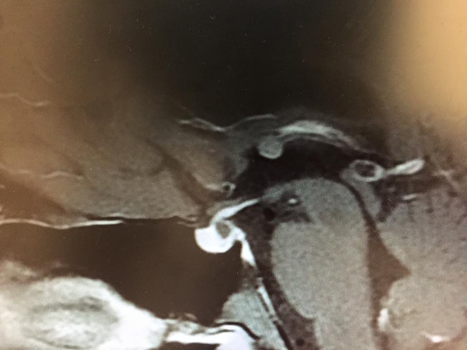
This is a mid sagital plane slice thru the pituitary. The whitest structure in the center of the image is the pituitary gland. In the upper right side of the image (which would be upper posterior area) has what looks like a hole. This is a small Rathke’s cyst. If you considered that the center of a clock and moved up to about one o’clock, you see a rounded structure with a little white wisp that seems like a tail. That is a colloid cyst of the third ventricle. If you consider that the center of a clock and go to the four o’clock position there is a dark hole with a white ring around it. this is a pineal cyst. Thus, this patient has THREE cysts on the same slice. Fortunately, none of them require surgery and they have been stable in size for 5 years.
© 2015 – 2017, Pituitary World News. All rights reserved.