From Lewis S Blevins, Jr. MD – Pathologists are the mystery men and women behind the scenes in your healthcare. You will never meet them. You might see their name on a report or two in your file. Or, perhaps on a bill for services rendered. They are, however, an integral part of your care for they firm up the clinical impressions and provisional diagnoses that are rendered based on patterns of presentation on history and physical, laboratory tests, radiographs, etc. In other words, they provide the “proof” of the final anatomical diagnosis. They do so in one of two ways: looking at tissue removed from patients with their eyes and evaluating same under the microscope.
Pathologists are often called upon by surgeons to provide a provisional diagnosis based on “frozen section” analysis of tissue resected at surgery. This technique is not always necessary in routine cases of suspected pituitary adenoma and may be more useful in those patients where it is unclear as to what might be the diagnosis and especially when certain diagnoses may lead to a limited rather than a complete resection. For example, patients with Germinoma, sarcoidosis, or one of the inflammatory conditions of the pituitary gland require biopsy and not total removal of their lesions. Frozen section analysis is performed by taking fresh tissue removed at the time of surgery, freezing it rapidly, slicing through it with a special knife called a microtome that can cut through frozen tissue, and then preparing slides that the pathologist will review to determine if there is sufficient information on which to base a diagnosis. This process is fairly rapid and takes place while the surgeon is continuing to perform the operation. Feedback is provided that guides the surgeon as to the extent of resection required.
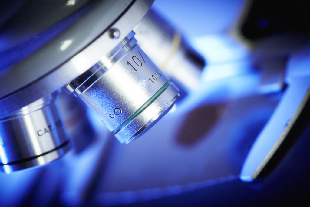
The most useful examinations of resected tissue take place about a week later. Tissue removed at surgery is placed in formalin and “fixed” in preparation for additional study. Then, paraffin wax is infiltrated into the tissues to replace fat and other oil-based substances. They wax blocks are then cut with the microtome. Microscope slides are prepared by placing these thin slices on the slide and fixing them to it so they do not move around. Next, the tissue is stained with a number of different chemicals to provide information about cell type, cell size, etc. In many cases, antibodies are used in immunoperoxidase “stains” to detect the presence of certain proteins, including pituitary hormones, and other cell markers.
The first order of business for the pathologist is to determine if there is sufficient material for examination. Next, the doctor will determine the type of tumor or other disease process that has affected the pituitary necessitating surgery. This is done by evaluating every aspect of the tissue presented relative to known information about the visual appearance of differing disease processes. Pituitary adenomas have certain characteristics, such as the pattern of the distribution and appearance of cells comprising the lesion, the loss of the reticulin architecture, etc. Once the tissue is identified as pituitary adenoma, immunoperoxidase stains allow the pathologist to determine what, if any, hormones were produced by the tumor. This is important as it provides confirmation of clinical syndromes of hormone excess such as acromegaly, hyperprolactinemia, Cushing’s syndrome, and hyperthyroidism. It also tells whether there is silent hormone production but not secretion by the tumor as is often the case in patients with gonadotropin adenomas Who were, prior to surgery, thought to have nonfunctioning tumors.. Further, about 40% of all ACTH producing tumors are silent and present as “nonfunctioning” pituitary adenoma’s and these can be identified as well. The same scenario occurs in a small number of patients with GH producing adenomas that do not secrete and therefore do not cause a clinical syndrome of acromegaly. Interestingly, some of these clinically silent hormone-producing adenomas can later become clinically apparent cause hormone excess related to the type of hormone they secrete. Special stains can also be performed to look at the mitotic index of tumors which reflects the aggressive nature of a particular adenoma. This is useful because not all aggressive tumors display mitoses, or evidence of cell division, at the time of surgery. Ki-67 is the marker used to assess the “proliferative index.” p53 Immunostains evaluate for the Protane P 53 which is a market for more aggressive tumors. When tumors have a high proliferative index and are positive for P 53 they are said to be “atypical adenomas.” We usually treat these as more aggressive lesions. Pituitary carcinoma is a diagnosis reserved for those lesions that extend to other areas of the brain by discontinuous spread or metastasize to other parts of the body including lymph nodes in the neck, the liver, the spinal cord, etc. The pathologist cannot make a diagnosis of pituitary carcinoma by histological examination of resected tissue. If your doctor tells you “your tumor was benign…there was no cancer” Here she is not providing you any useful information. What you want to know is was The tumor a traditional adenoma or an atypical adenoma. Further, you want to know whether there was any evidence of hormone production whether or not a hormone was secreted in excess. Frankly, the histologic diagnosis is best correlated with the MRI findings and the overall clinical course. Together, these bits of information are used to render a treatment plan.
Worthy of mention is the fact that patients who have Cushing’s disease will have a characteristic perinuclear clearing and ground glass appearance in the cells in the normal normal pituitary that produce ACTH and this is referred to as Crooke’s hyaline change. The lack of this finding in pituitary tissue from patients thought to have Cushing’s disease is, as far as I’m concerned, evidence against a diagnosis of Cushing’s disease.
Some centers, and especially smaller hospitals, do not perform all of the standard test to examine pituitary adenomas. It may be useful to send your pituitary tumor slides or tissue block to a pituitary center of excellence for evaluation if they do not do things such as immunochemistry, Mib-1 labeling, and p53 studies.
Of course, pituitary adenomas are not the only tumors that affect the hypothalamic-pituitary unit. Pathologists distinguish between tumoral and other non-tumoral conditions based on the patterns of changes to normal tissues, type of cells present, connective tissue changes, inflammatory changes, and other features.
Whenever a pituitary disorder recurs, and repeat surgery is needed, it is often useful to compare the newly acquired tissue to the previously acquired tissue. This is necessary because, rarely, patients have a second and different type of tumor. For example, 2% of patients with a pituitary tumor will have more than one adenoma. The first adenoma may present years apart from the second one.
Finally, patients often want to obtain copies of their MRI reports. We suggest that you also ask for copies of your pathology reports and file them for safekeeping. This is especially true in the event you plan to move or otherwise change healthcare teams.
© 2014 – 2024, J D Faccinetti. All rights reserved.